

Cartilage Grows In Two Ways
Cartilage, Bone & Ossification: Cartilage
What is cartilage for?
- to class the supporting framework of some organs, such equally the walls of airways (olfactory organ, trachea, larynx and bronchi), where it prevents airway plummet.
- to class the articulating surfaces of basic, and
- to class the template for the growth and development of long bones, and most of the rest of the fetal skeleton (gradually replaced by bone). In children, the cartilaginous plates at the ends of long bones can be seen on X-rays. These templates disappear when adults reach their full pinnacle.
Constituents of cartilage.
Cartilage is a strong, flexible and semi-rigid supporting tissue. It can withstand pinch forces, and yet it tin bend. It is made upwards of cells called chondroblasts and chondrocytes, (chondro - cartilage) and extracellular matrix, made up virtually 10% aggrecan, 75% water, and a mix of collagen fibres and other constituents.
In that location are three types of cartilage, which differ mostly in the type of fibres they contain.
Hyaline: Most common - has a glassy appearance (hyalos - is greek for drinking glass).
Fibrocartilage: tendon insertions and invertebral discs: reinforced with parallel bundles of collagen fibres.
Rubberband cartilage: external ear and epiglottis: flexible and resilient - has rubberband fibres too equally collagen fibres.
Click here to find out more than virtually these three types.
This diagram shows the structure of an aggregate of Aggrecan (another proper noun for the aggregating GAG chondroitin sulphate).
Aggrecan is only found in cartilage. The aggregates contain upwardly to 100 molecules of chondroitin sulphate (shown in purple) are bound to 1 molecule of hyaluronic acrid (shown in ruddy).
Importance of Extracellular matrix
The extracellular matrix soaks up water, like a sponge, (osmotic effect) due to the properties of the GAG: aggrecan, producing the high water content (75%).
The collagen fibres form a network which has a very high tensile forcefulness, and which entraps the aggrecan molecules.
When y'all stand or walk, the weight of your body is supported past the cartilagenous ends of the long bones. In this state your weight compresses the cartilage, literally squeezing the water out, until the force produced by the osmotic swelling is equal to the compressive force generated by your weight.
Cells
The extracellular matrix of cartilage is secreted by chondroblasts, (chondro = cartilage), which are found in the outer roofing layer of cartilage. As the chondroblasts secrete matrix and fibres, they become trapped within it, and mature into cells called chondrocytes. (See diagram contrary)
In growing cartilage, the chondrocytes tin divide, and the girl cells remain close together in groups, forming a 'nest' of 2-4 cells. The matrix enclosed compartments that they sit in are called lacunae. (lacunae = little lakes/small pits). The active chondrocytes are large secretory cells with basophilic cytoplasm considering they have lots of rough endoplasmic reticulum. Older chondrocytes contain fat droplets.
(Fixation of cartilage usually causes some shrinkage between the cell border and the lacunar wall, so that these lacunae look more prominent in stock-still tissue.)
The surface of most cartilage is covered past a layer of dumbo irregular connective tissue called the perichondrium (peri = around). The outer layer of the perichondrium contains collagen producing fibroblasts, and the inner layer contains chondroblasts.
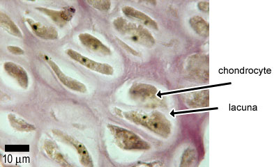
This is a photograph of a section of hyaline cartilage, showing the chondrocytes trapped in lacunae in the matrix
Growth and nourishment of cartilage:
Different other connective tissues cartilage is avascular (similar epithelia). Cartilage is nourished past long range diffusion from nearby capillaries in the perichondrium. Therefore, cartilage can never become very thick, as diffusion would not exist sufficient to supply the cartilage with nutrients and oxygen. (This is in contrast to bone, because os has a very expert claret supply).
Cartilage can grow in two ways: Interstitial growth - chondrocytes grow and divide and lay down more matrix within the existing cartilage. This mainly happens during childhood and adolescence. Appositional growth - new surface layers of matrix are added to the pre-existing matrix by new chondroblasts from the perichondrium.

Cartilage Grows In Two Ways,
Source: https://www.histology.leeds.ac.uk/bone/cartilage.php
Posted by: harrisfooke1944.blogspot.com
0 Response to "Cartilage Grows In Two Ways"
Post a Comment